
Products
Orthodontics


FACAD CEPHALOMETRIC SOFTWARE
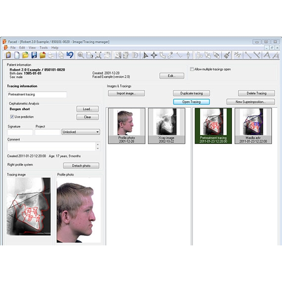
Facad® is a software program used for orthodontic tracing, cephalometric analysis, and visual diagnostic imaging, as well as for treatment planning with soft tissue profile prediction for both orthodontics and maxillo-facial surgery. This program is meant to be used by orthodontists and orthofacial surgeons.
Facad is EC certified (CE marked) according to the Directive 93/42/EEC on Medical Devices, annex II (MDD). The software is developed by the Swedish company Ilexis AB in co-operation with the maxillofacial unit at the University Hospital in Linköping, Sweden.Facad has been in clinical use for more than 20 years and is available as a state-of-the-art PC program for Windows 7/Vista/XP
FEATURES
- Import of digital images
- JPEG, TIFF, BMP, DICOM
- Scanned x-ray films
- Digital x-ray images
- Cephalograms
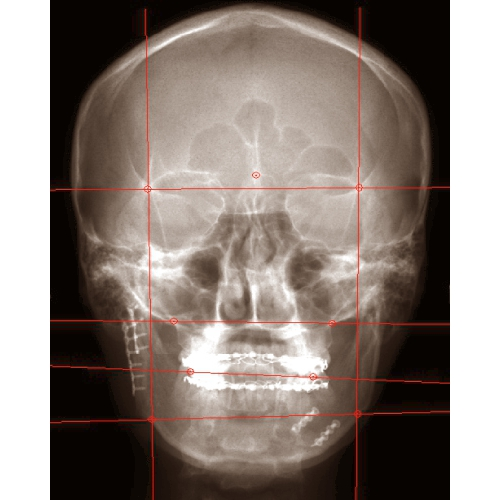
- Frontal x-rays
- Panoramic x-rays
- Facial photos
- Lateral
- Frontal
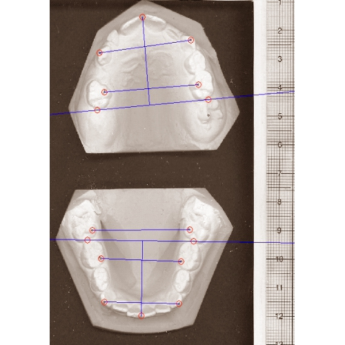
- Cast models (2-D)
- Photographed
- Scanned models
- Other images
- Paste copied image data from the Windows desktop
- Connection with software programs for digital x-ray imaging, patient management systems, or PACS, see list of software
- Multiple language support
- English, Swedish, Norwegian, Danish, Finnish, French, Spanish, Dutch, Polish, and Slovenian.
- Visual overview (with image thumbnails) and management of a patient's images, tracings, treatment plans, and superimpositions.
- Image calibration
- Real world calibration, using the system ruler in the x-ray image
- Enter the resolution (mm/pixel) of the x-ray detector
- Enter the DPI (dots per inch) resolution of a flatbed image scanner (for scanned x-ray film)
- Compensation for x-ray enlargement factors
- Image functions
- Zoom
- Enlarge/reduce in steps
- Enlarge/reduce continuously
- Brightness and contrast
- Adjust brightness and contrast
- Optimise brightness and contrast in a specific image area
- Invert an image
- Rotate an image 90°/180°, flip up side down, or mirror left/right
- Free image rotation
- Zoom
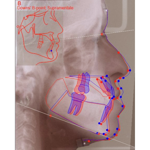
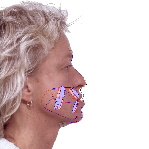
- Tracing
- Place landmarks
- A guided process with visual placement guide for landmarks
- Automatic positioning of the mouse cursor
- Place only the landmarks necessary for the chosen analysis
- Adjust the position of landmarks
- Place/adjust landmarks with high precision
- Automatic placement of incisors
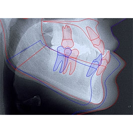
- Place teeth using ready-made templates
- Adjust the position, size and slope of teeth
- Draw hard tissue (maxilla, mandible, and other anatomical structures) using ready-made templates
- Adjust the hard tissue structures
- Draw the soft tissue profile line using a template and automatic structure recognition
- Adjust the soft tissue profile line
- Trace on either right or left cephalograms.
- Undo any actions easily, to correct for user mistakes
- Place landmarks
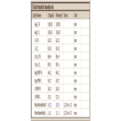
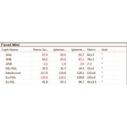
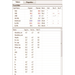
- Cephalometric analysis
- Predefined standard analyses
- lateral analyses
- frontal (PA) analyses
- cast model analyses
- Modify and custom-design your own analyses
- Adjust norm intervals for selected measurements
- Build your own analyses from a measurement library
- Flexible graphic presentation of measurements, values, and cephalometric lines
- Print images, tracings, analysis results, and combined reports
- Export images to image files
- Save analysis results to text files
- Copy images, tracings, and analysis results to the Windows clipboard
- Export Facad patients to e.g. a memory stick
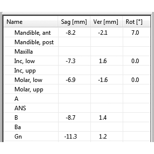
- Planned movement and rotation of teeth, maxilla, and mandible - with high precision, interactively or by entering numeric movement values
- Immediate numeric presentation of planned movement
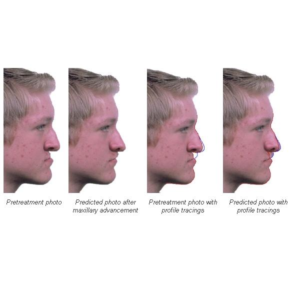
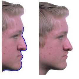
- Immediate soft tissue prediction
- Simulate effects from automatic rotation of the mandible
- Split hard tissue segments
- Simulate the use of distractors
- Predicted photo as a result from treatment planning
- Flexible model for soft tissue movements
- Multiple planning suggestions
- Superimpose a lateral x-ray with a profile photo with see-through effect
- Superimpose two x-ray images with see-through effect
- Superimpose 2-5 tracings simultaneously
- Present analysis values from all superimposed tracings simultaneously
- Superimpose tracings before/during/after treatment
- Superimpose tracings during growth
- Superimpose a treatment plan with a post-treatment tracing, i.e. compare a treatment plan with the actual result
- Measure differences (distances) between tracings in a superimposition
- Several methods for alignment of included tracings:
- Fixate tracings at a reference marker, rotation by a reference line
- Best match using 2 or more reference markers
- Manual alignment, use anatomical structures
- Perform the Structural Method by prof. Arne Björk (with implant lines)
- Complete Reference Manual
- User's Guide in several languages
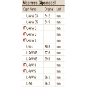
- Well documented standard analyses
- Measurement library documentation
- Release Notes
Highlighted Features
- Facad is a very powerful, flexible & easy to use software program used for Orthodontic tracing, cephalometric analysis and visual diagonostic imaging as well as for treatment planning with soft tissue profile prediction for both orthodontics and maxillo facial surgery.
- It integrates and readily adaptable with other softwares for receiving patient data and dental images.
- Facad enables a quick and easy method which involves a guide and automatic positioning of cursor to place anatomic landmarks directly on the digital Xray image with great accuracy.
- Teeth,Hard tissue segments such as maxilla & mandible and other anatomical structures are placed/ drawn on X-ray using ready made templates. There are about 28 Cephalometric analyses,4 cast model analyses & 2 frontal analysis.
- Creation of own Composite analysis is also possible. Superimposition of different analysis and treatment planning can be made and measurement of the analysis is readily available.
- Blending of X-ray and profile photo simultaneously with see through effect is possible.
- Reports of the patient with tracing, analysis, profile and predicted photo can be generated.